

")
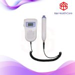
Vascular Doppler is a device that ultrasonic control of vessels using different MHz values. It provides a direct evaluation of how blood flows to the arteries in real time using vascular Doppler. At the same time, narrowing of arteries, blockages and blood clots can be detected during Vascular Doppler use. The course of arterial diseases is easily monitored. Vascular doppler allows a direct look at how blood flows to the arteries in real time. In doing so, narrowing of the arteries, blockages and even blood clots can be detected. The course of arterial disease is easily monitored. A vascular doppler sonography is called vascular ultrasound. The ultrasound test uses high-frequency sound waves to generate an image of the area under investigation. This is done with a transducer printed against the skin.
Vascular Doppler Working Principle
Vascular Doppler uses sound waves at different frequencies to generate an image of the area under investigation. A transducer printed against the skin helps to create this image. The sound waves sent by the transducer are reflected back to save the image. This image is instantly transmitted to the monitor and saved. For vascular Doppler sonography, these sound waves are concentrated on the patient’s heart and blood vessels.
A clean water-based gel is applied to the examined areas to create a better contact surface. In some cases, it may be necessary to apply blood pressure cuffs to the patient’s arms and legs to see the values in the vessels being examined. The transducer sends the sound waves and the waves record the reflected image back. This image is instantly transmitted to a video monitor for visualization and can be recorded on video. For vascular Doppler sonography, the sound waves are concentrated on the patient’s heart and blood vessels. While almost all blood vessels can be examined with such sonograms, the doctor is usually only interested in a few large vessels and vessels. The most popular areas are the legs, abdomen and head and neck arteries and vessels.